Products

SUTTER INSTRUMENT, USA

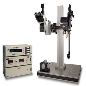
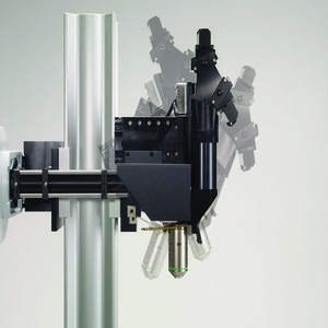
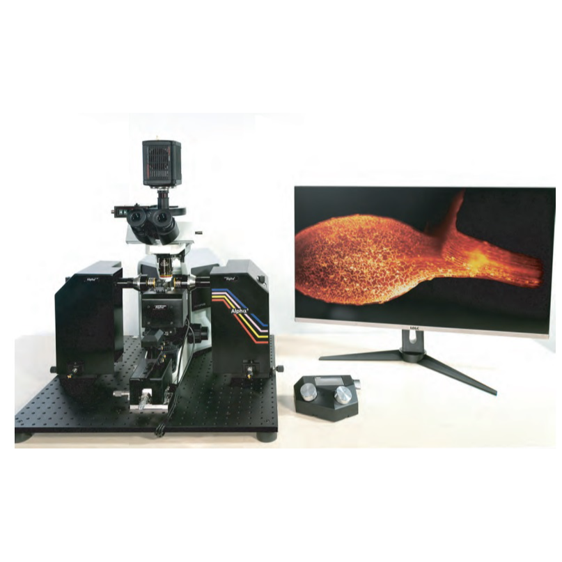
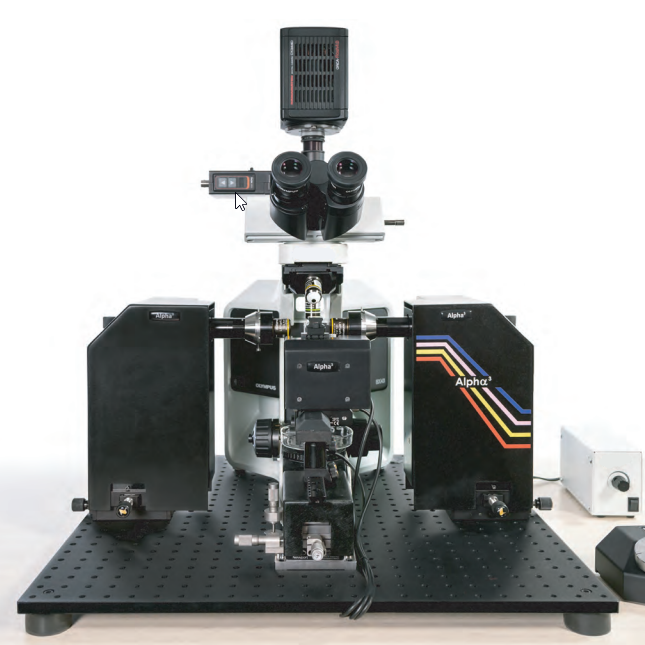
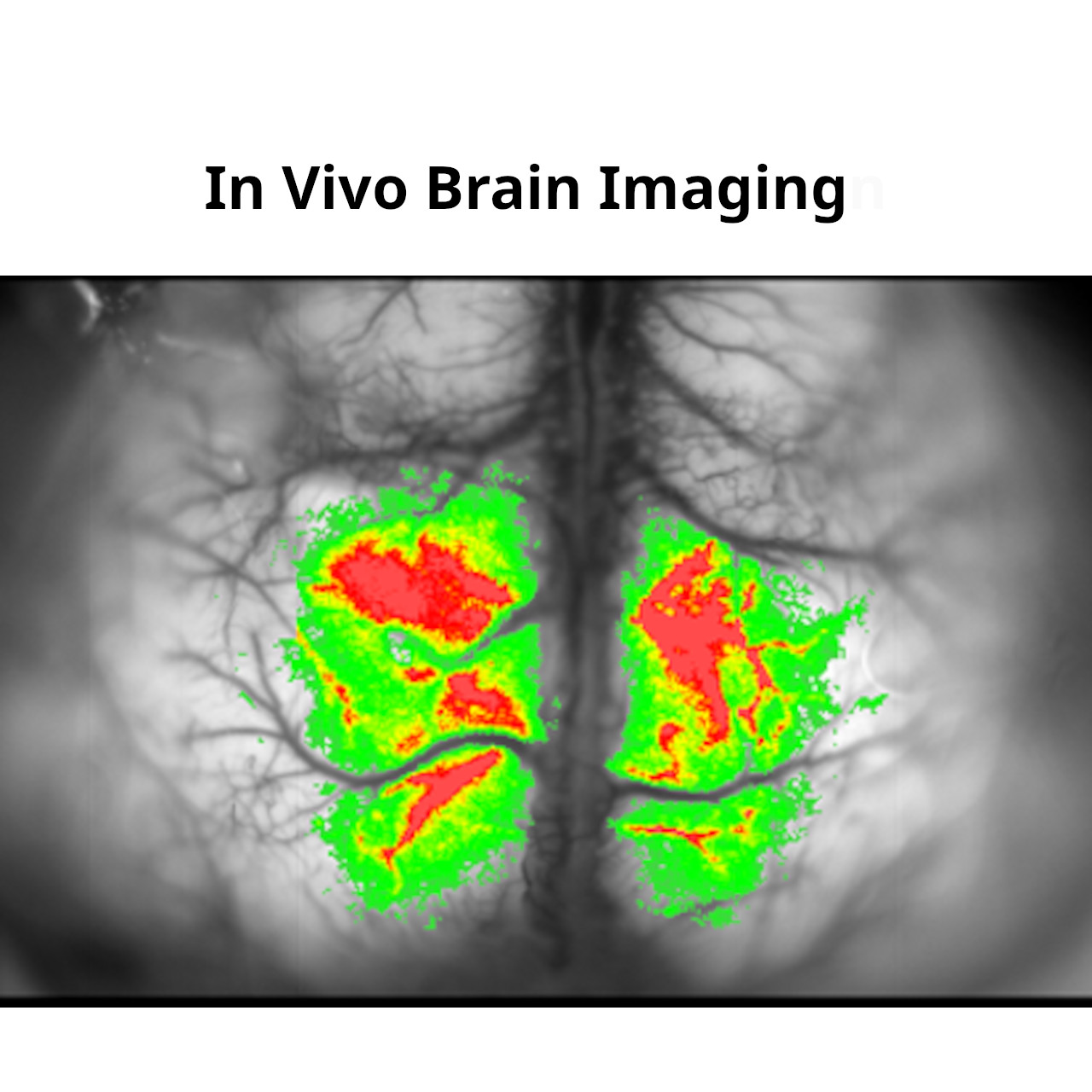
MOM® Movable Objective Microscope®

    FEATURES
  • Objective moves 22mm in X, Y, and Z
  • Objective rotates about optical axis for imaging of non-horizontal surfaces and volumes
  • Customizable open platform design
  • Cambridge Technology conventional or resonant XY scanners
  • Two or four channel detector system with Hamamatsu PMTs and preamplifiers
  • Sutter PS-2/ PS-2LV dual channel PMT power supply
  • Two- or three-photon compatible scan lens and tube lens
  • New Light Block keeps visual stimuli, photostimuli, and ambient light out of detector path
  • National Instruments and Vidrio Technologies data acquisition systems
    APPLICATIONS
  • In vivo two-photon imaging
  • In vivo three-photon imaging
  • Electrophysiological recording and imaging (culture, large in vivo preparations, etc.)
  • Non-horizontal surface microscopy
  • Simultaneous retinal stimulation and two-photon microscopy
  • Whole animal imaging
  • Immunology
  • Embryology

TOMOCUBE INC., SOUTH KOREA

HT-1 : Holotomography microscope to view living cells non-invasive, real time quantitative data

HT-2 : Holotomography microscopy with 3D Fluorecence imaging capability 

HT-X1 : High-Performance Holotomography Microscope for imaging at scale

  • High resolution, high contrast, and high sensitivity
  • Large captured fields
  • Programmable multi-well plate imaging
  • Laser autofocus for well-to-well reproducibility
  • Low noise, speckle-free imaging with no calibration step
  • Bioimaging made quantitative
  • Integrated incubation for long term timelapse
  • Integrated 4-channel FL light engine (Blue, Green, Red, Far red)
  • Correlative holotomography with 3D fluorescence
  • True multi-dimensional imaging across time, space and modality

Applications :- 

  • Cell biology : Lipid imaging, Neuron imaging, Single cell study, Cancer biology
  • Experimental Laboratory : Ob/Gyn, Infectious diseases, Immunology, Hematology
  • Microorganism : Plankton, Protozoa, Bacteria
  • Other applications include : Pollen studies, stem cells, nanotechnology, etc.


IVIM TECHNOLOGY INC., SOUTH KOREA

The world’s first All-in-One intravital confocal/two-photon microscopy system (IVM-C/M/CM/CMS/MS2) microscopy platform enables dynamic 3D cellular-level imaging of various biological processes in living animals in vivo.

In vivo visualizations of gene expression, protein activity, cell trafficking, cell-cell / cell-microenvironment interaction.

Explore complex dynamic behaviors of numerous cells inside a living body

Applications

  • Live Cell Intravital Imaging
  • Repetitive Intravital Imaging
  • In vivo Drug Efficacy Monitoring
  • In vivo Drug Delivery Monitoring
  • Ex Vivo Imaging

PHASE HOLOGRAPHIC IMAGING (PHI), Sweden

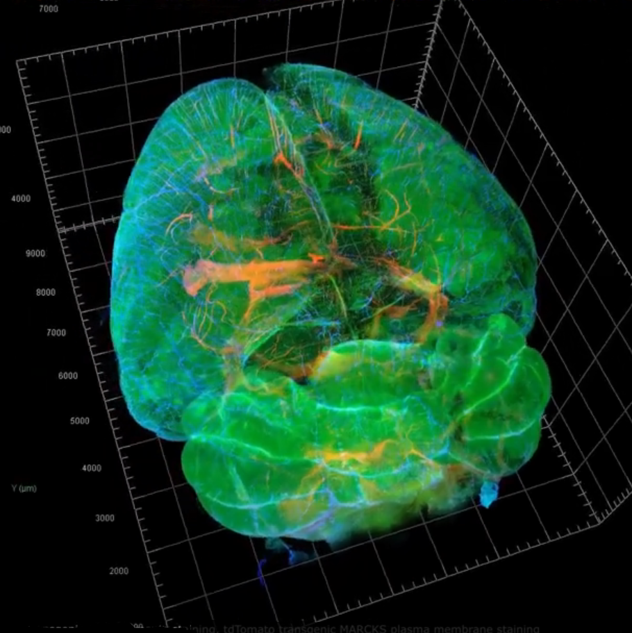
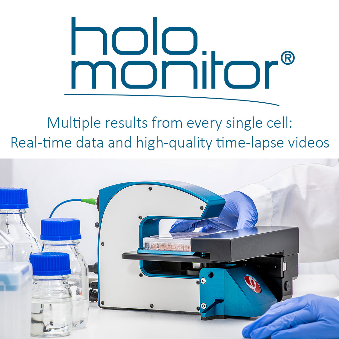
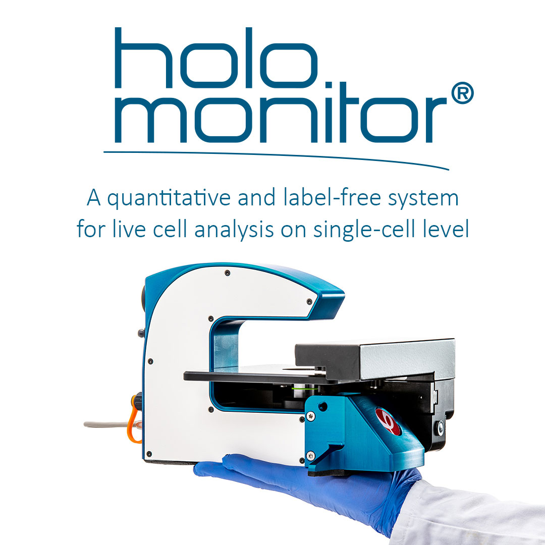
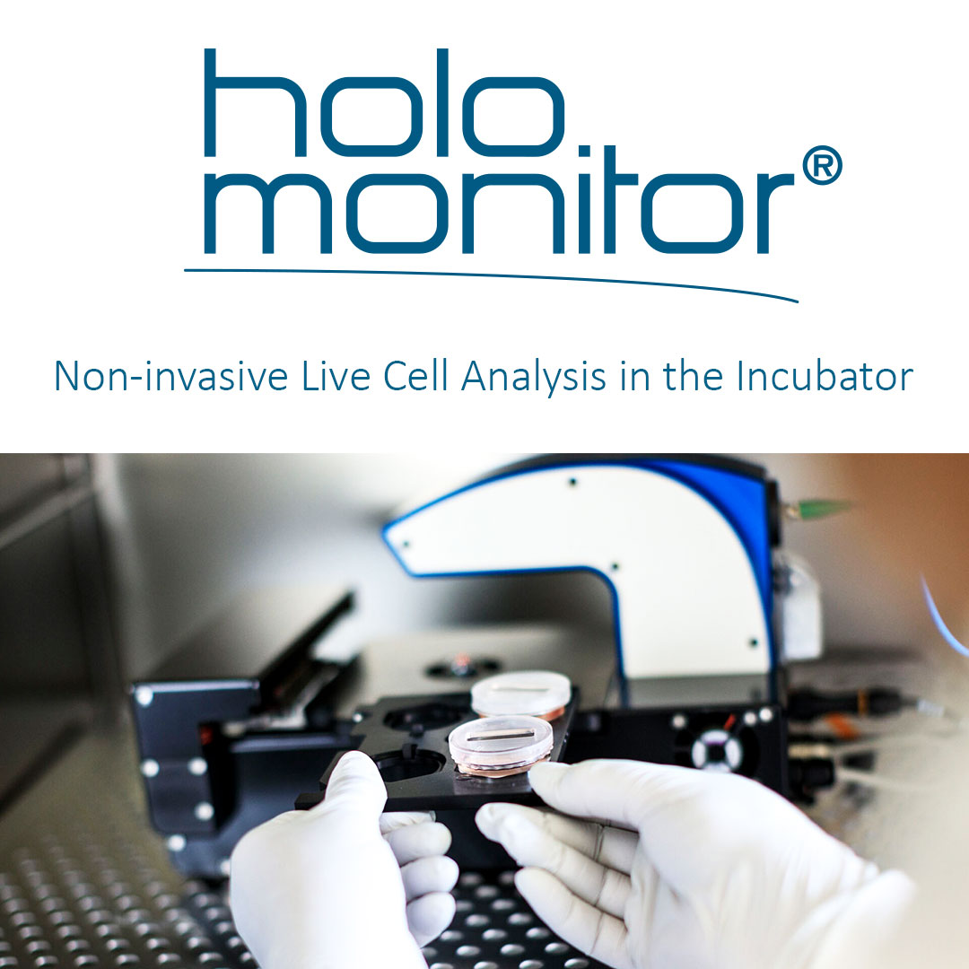
HoloMonitor holographic microscopy (incubator -suited) to image cells in real-time without cellular labels or stains.
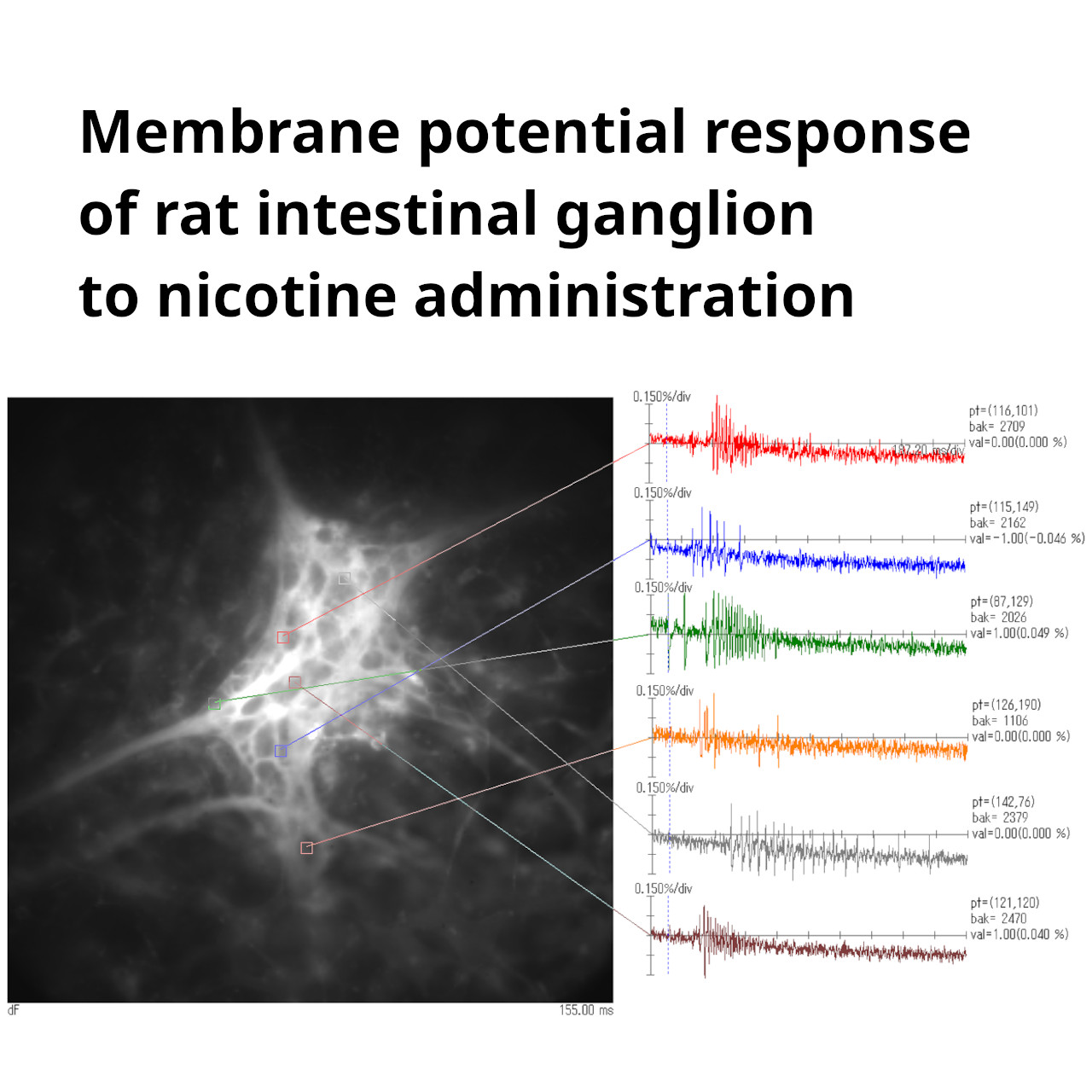
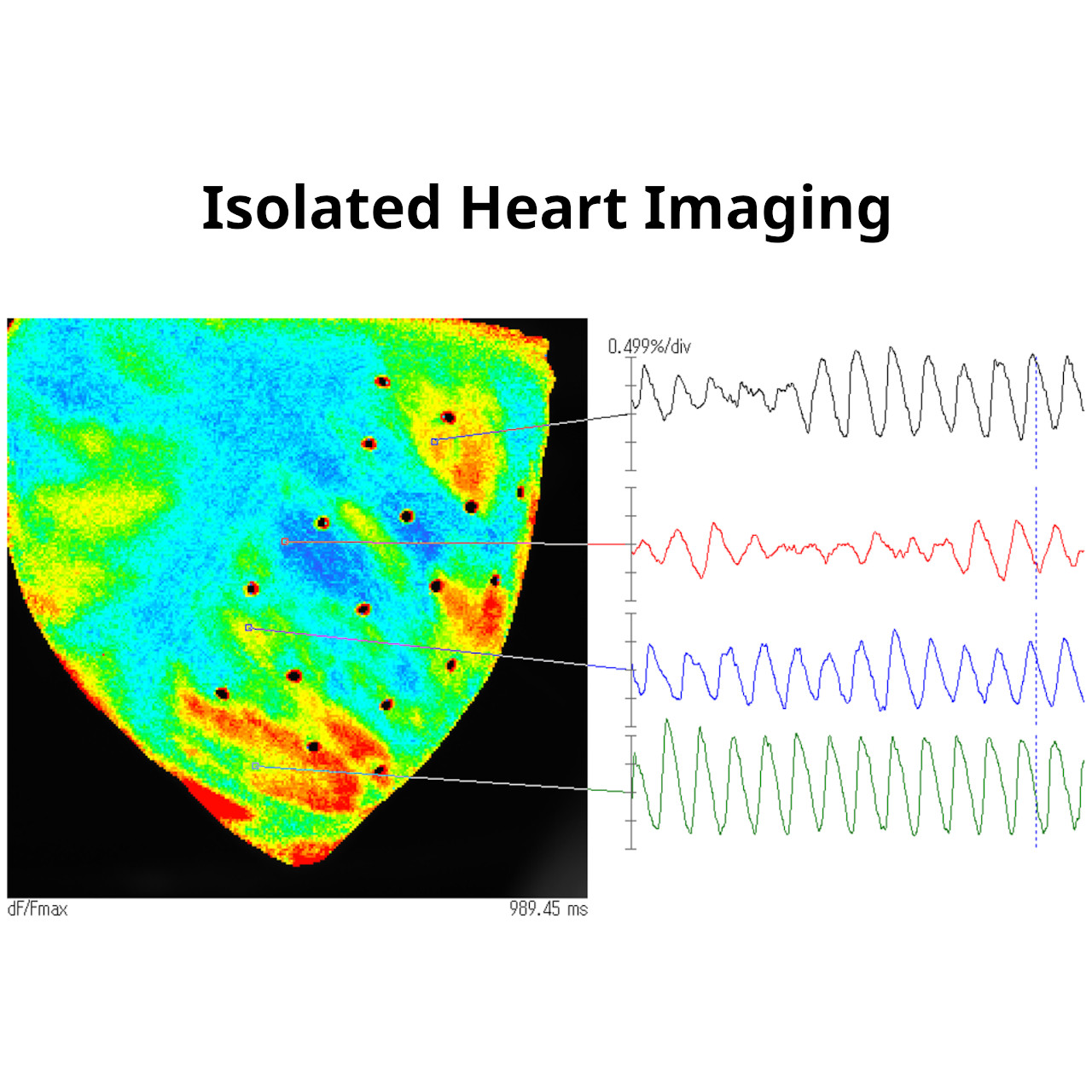
Applications :

  • Kinetic Cell Morphology (with data on 30+ parameters)
  • Automated Single Cell Tracking & Analysis
  • Kinetic Cell Motility & Migration
  • Wound Healing (Scratch assay) | Chemotaxis
  • Kinetic Cell Proliferation
  • Cell Growth | Cell Division | Cell Cycle
  • Cell Differentiation
  • Kinetic Drug Dose-Response
  • Cytotoxicity | Cell Death
  • Cell Counter
  • Cell Quality Control (QC)

FEMTONICS LTD, HUNGARY

FEMTO3D Atlas combines an intoxicating blend of high-tech science, engineering, refinement in 3D measurements.

FEATURES AND BENEFITS
  • Available both as a 3D imaging extension for various types of existing microscopes and as an individual system as well
  • Simultaneous read out of neural activity on both the somatic and dendritic scales
  • Increased measurement speed and signal-to-noise ratio by several orders of magnitude
  • 3D Network imaging of over 2000 soma distributed in 3D
  • Imaging of dendritic branches through layers
  • Simultaneous imaging of over thousands of spines
  • Unique, flexible scanning methods supporting in vivo and in vitro measurements
  • In vivo real-time 3D motion correction 
  • High-speed 3D random-access ROI scanning: scanning speed up to 30 kHz to any points in 3D
  • High-speed raster scanning: 40 fps at 510×510, and even 3000 fps at reduced FOV
  • Improved excitation efficiency for large 3D scanning volume with GECIs
  • Automatic wavelength tuning between 750 – 1050 nm
  • Functional imaging correlated with electrophysiological data
  • Calcium imaging during behavior
  • 3D random-access photo stimulation for optogenetics

The FEMTOSmart series is the next step in classical two-photon microscopy at Femtonics, fully customizable two-photon microscopes.

FEATURES AND BENEFITS

  • Elevated, column-based body
  • XYZ positioning
  • Tilting objective
  • High level of modularity
  • Galvanometric, resonant, or dual scanning possibilities
  • Galvo Scanner, focusing on regions-of-interest
  • Resonant Scanner, fast frame and volume scanning
  • Dual Scanner, advantages of Galvo and Resonant microscopes combined
  • Travelling detector system
  • For in vivo studies
  • High number of optional modules
  • Patented imaging technologies
  • Flexible scanning methods
  • Maximal photon collection

SCIMEDIA LTD, USA - BRAINVISION INC. JAPAN

  • Imaging system: Imaging systems that can capture small changes in fluorescence light intensity ( membrane potential, calcium ion concentration) emitted from biological samples.
  • Data analysis software for optical imaging
  • Fluorescence Macroscope : A tandem-lens macroscope fluorescence developed for membrane potential imaging and calcium imaging.
  • Light source: An LED light source that has both high light intensity and stability for excitation of fluorescence imaging.


PHI OPTICS INC., USA

PHI OPTICS  introduces a novel technology – Spatial Light Interference Microscopy (SLIM) – that provides label-free, quantitative, and real-time imaging of live cells and tissues. SLIM is an add-on to all major brand optical microscopes and seamlessly overlays with fluorescence for multichannel imaging. SLIM modules connect to the microscope like a camera and use the white-light illumination source of the microscope

PHI OPTICS  technology provides: 

Real-time quantitative phase imaging and display 

Non-invasive, label-free cell and tissue imaging on broad time scales (seconds to weeks) 

Programmed 2D and 3D scanning for large area 2D SLIM maps and 3D SLIM images 

Seamless overlay with other microscopy channels 

Quick and easy segmentation of cells 

Applications include: 

Cell growth 

Cell dynamics 

3D tomography 

Neuroscience 

Blood testing 

Tissue imaging

KA IMAGING INC, USA

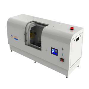
inCiTe – World’s first benchtop propagation-based phase contrast 3D X-ray micro-CT

  • BrillianSe™ Amorphous Selenium (a-Se) detector 
  • High spatial resolution and detection efficiency
  • Phase contrast technology for superior contrast 
  • Excellent visualization of biomaterials, polymer composites and low-density materials

Reveal35C - World’s first portable single-shot dual-energy portable x-Ray detector

  • Single shot, dual energy detector
  • Triple stacked layer design - 3 images in 1 single exposure
  • Portable - wireless connectivity
  • Single exposure, no motion artifacts 
  • Spectral information & color images
  • Advanced material separation 

CELLSORTER COMPANY FOR INNOVATIONS - HUNGARY

Robotic Isolation Technology

CellSorter device with valve control

CellSorter piezoelectric sorting head

Full CellSorter instrument with Valve Control

ENQUIRY ?